Summary
The severe neurological symptoms seen in children with PCH2A are caused by how the disease affects the brain. MRI allows us to visualize these abnormalities more precisely and thus gain important insights into the disease.
The aim of this work package is therefore to investigate the long-term progression of the disease on MRI, as well as to use new MRI technology to obtain more detailed information on networks and structures in the brain. In the future, our data is intended to help with disease prognosis in affected children, with the development of cell-based investigative approaches (organoids), and possibly one day with the assessment of therapeutic applications.
Background Information
Magnetic resonance imaging (MRI) is an essential part in the diagnosis of PCH2A. The stunted growth of the cerebellum and additional effects of the disease on the brain can be visualized. In older children, important information about how the disease progresses can also be obtained.
However, this poses challenges for research into PCH2A:
- As PCH2A is very rare, the researchers working at each medical center have only a few data sets available to them
- Often just the most important data is recorded to confirm the diagnosis, which does not utilize the full potential of MRI
Together, these factors are the reason why the existing knowledge about the effects of PCH2A on the brain is almost exclusively available for infancy and is limited to basic data, such as the size of the cerebellum. There is little known about the long-term effects of the disease on the brain. In addition, the majority of studies published so far only refer to a small number of cases, which detracts from the significance of the findings.
Objectives of the Research Project
This work package has two objectives:
The collection and evaluation of existing MRI data: With the support of the parents’ initiative PCH2A-Familie e.V., we were able to collect existing MRIs from a large number of affected children. The age range extends from children still in the womb to teenage patients.
This unique dataset allows us to extend the existing knowledge of PCH2A to older children. This allows us to answer questions such as:
- Up to what age does the brain continue to grow? Does the size then remain constant or does it decrease?
- Are all areas of the brain affected to the same extent, or are there regional differences?
- Is there a correlation between the size of the brain and the child’s cognitive and motor development?
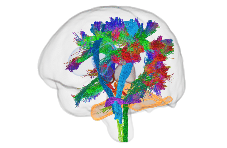
Deepen our understanding with modern imaging and analysis techniques: MRI methods are constantly being advanced. Modern techniques enable a detailed study of nerve cells and their connections, but also require repeated MRI scans. This data is collected as part of check-up examinations; we use it to examine the connections between the cerebellum and cerebrum, the connections within the cognitive and motor networks of the brain, and the effects of PCH2A on the nerve cells in different areas of the brain.
Outlook
Based on our statistics on the long-term progression of the disease, both families and healthcare professionals can better predict how the disease will affect individual children in the long term. Deviations from the normal course of the disease can also be recognized earlier and treated if necessary.
Researchers in the “Organoids” work package can also use our data to assess the extent to which their cell models reflect reality. Should it turn out that certain brain structures are affected particularly strongly in PCH2A, researchers could focus on these areas in order to better understand the cellular processes of the genetic defect.
Finally, if a therapeutic approach for PCH2A were to be developed one day, the development of treated children could be compared with our baseline data to assess its effectiveness.
Conclusion
MRI can be used to obtain important information about the effects of PCH2A on the brains of patients without causing pain or exposing them to radiation. On the one hand, we are pooling the data already collected in order to provide the information contained therein to families, treating physicians and researchers in the best possible way; on the other hand, we are using modern methods to analyze new MRI data, revealing differentiated information about the effects of PCH2A on brain networks and structures.


